45 cell membrane diagram with labels
Labeled Plant Cell With Diagrams | Science Trends The parts of a plant cell include the cell wall, the cell membrane, the cytoskeleton or cytoplasm, the nucleus, the Golgi body, the mitochondria, the peroxisome's, the vacuoles, ribosomes, and the endoplasmic reticulum. Parts Of A Plant Cell The Cell Wall Let's start from the outside and work our way inwards. Cell Membrane With Labels Labeled : Functions and Diagram evmestycor: cell membrane diagram (Virgie Goodwin) There is a printable worksheet available for download here so you can take the quiz with pen and paper. In a plant cell, the cell wall is made up of cellulose, hemicellulose, and proteins while in a fungal cell, it is composed of chitin.
Cell Membrane (Plasma Membrane) - Genome.gov Definition. …. The cell membrane, also called the plasma membrane, is found in all cells and separates the interior of the cell from the outside environment. The cell membrane consists of a lipid bilayer that is semipermeable. The cell membrane regulates the transport of materials entering and exiting the cell.

Cell membrane diagram with labels
CELL MEMBRANE LABEL Diagram | Quizlet Practice labeling the parts of the cell membrane Terms in this set (6) Channel Protein hole or tunnel that particles may pass through to go in / out of cell Marker protein identifies or labels the cell Receptor protein receives information Heads part of the phospholipid that loves water (hydrophili) - points to the most outside and inside of cell Plant Cells: Labelled Diagram, Definitions, and Structure - Research Tweet Plastids and Chloroplasts. Plants make their own food through photosynthesis. Plant cells have plastids, which animal cells don't. Plastids are organelles used to make and store needed compounds. Chloroplasts are the most important of plastids. They convert light energy from the sun into sugar and oxygen. The most exposed parts of the plants ... Structure of Membrane in Cells (With Diagram) - Biology Discussion Cell walls are formed through apposition, i.e., the wall material is deposited by the protoplast on the plasma membrane. The first cell wall (Primary wall) is formed during the cell growth phase. When the cell elongation is stopped the secondary cell wall, formation starts (Fig. 2.13).
Cell membrane diagram with labels. Labeled Diagram Of Cell Membrane : Prokaryotic Cell Structure Diagram ... The outer covering of the body cells, which maintains homeostatic condition between inside and outside of the cell is called cell membrane. Schematic diagram of a cell membrane membrane structure, cell structure,. It is made up of . Membrane proteins labeled vector illustration. Learn how to find cell towers near you. Cell Membrane Function and Structure - ThoughtCo The cell membrane (plasma membrane) is a thin semi-permeable membrane that surrounds the cytoplasm of a cell. Its function is to protect the integrity of the interior of the cell by allowing certain substances into the cell while keeping other substances out. Labeled Diagram Of Cell Membrane : Electron Micrograph The nucleus and mitochondria are two examples. Copy of labeling cell membrane labelled diagram. Some of the major parts of the plasma membrane are : Phospholipid bilayer · phospholipid bilayer ; It supports and helps maintain a cell's shape. 1)cell membrane 2)vacuole 3)nucleus 4)endoplasmic reticulum 5)mitochondria 6)golgi body. Cell Organelles- Definition, Structure, Functions, Diagram - Microbe Notes A cell wall is multilayered with a middle lamina, a primary cell wall, and a secondary cell wall. The middle lamina contains polysaccharides that provide adhesion and allow binding of the cells to one another. After the middle lamina is the primary cell wall which is composed of cellulose.
Labeling a cell membrane Diagram | Quizlet Start studying Labeling a cell membrane. Learn vocabulary, terms, and more with flashcards, games, and other study tools. Label the Cell Membrane - Labelled diagram - Wordwall Label the Cell Membrane - Labelled diagram channel protein, cholesterol, external cell environment, hydrophilic (water loving) part of phospholipid bilayer, peripheral protein. Basic Cell Membrane Label - Labelled diagram - Wordwall Integral Protein (channel), Peripheral Protein, Phosphate, Lipid, Hydrophilic, Hydrophobic, Glycoprotein. Plasma Membrane Function, Structure & Diagram - Study.com 2. property of the plasma membrane that allows some substances into the cell and keeps others out 4. main structural component of the plasma membrane 6. nonpolar part of a phospholipid 11. protein...
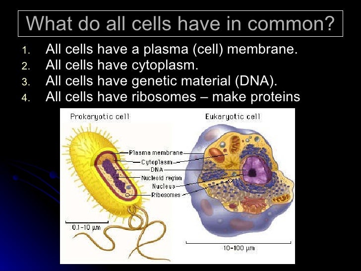
diagram of cell membrane labeled Plasma Membrane With Parts Labeled, Hydrophilic, Hydrophobic membrane protein transmembrane plasma hydrophobic hydrophilic proteins integral labeled parts peripheral cells k12 Root Hair Cell Parts Diagram (Trevor Mills) trevor liver thinglink The Plant Cell Is Like A Home.. PDF Cell Membrane Structure (1.3) - University of São Paulo Cell Membrane Structure (1.3) IB Diploma Biology Essential idea: The structure of biological ... When drawing a diagram of a phospholipid this is a good example which shows all the key features 1.3.1 Phospholipids form bilayers in water due to the amphipathic ... • Labels clearly written • (Scale bar if appropriate) ... Cell: Structure and Functions (With Diagram) - Biology Discussion 1. Eukaryotes are sophisticated cells with a well defined nucleus and cell organelles. 2. The cells are comparatively larger in size (10-100 μm). 3. Unicellular to multicellular in nature and evolved ~1 billion years ago. 4. The cell membrane is semipermeable and flexible. 5. Animal Cell Diagram with Label and Explanation: Cell ... - Collegedunia The cell membrane is the thin semi-permeable layer surrounding the cell; its main functionality is to protect the cell. It has hair-like structures cilia and flagella on it. The cell membrane controls the entry and exit of nutrients into the animal cell. Cytoplasm
Cell membrane with labeled educational structure scheme vector ... 2. Editable Vector .EPS-10 file. 3. High-resolution JPG image. Use for everything except reselling item itself. Description: Cell membrane with labeled educational structure scheme vector illustration. Anatomical closeup drawing with cross section element. Carbohydrate, globular protein or cholesterol location visualization.
Diagram of a cell membrane with labels | NIST Essential Biological FunctionsImmune response, Cell metabolism, Neurotransmission, Photosynthesis, Cell adherence, Cell growth and differentiationPotential Commercial ApplicationsDrug response monitoring, Chemical manufacturing, Biosensing, Energy conversion, Tissue engineering ... Diagram of a cell membrane with labels. Appears In. Biology in ...
The Cell Diagram Label Plant - dim.uds.fr.it Search: Label The Plant Cell Diagram. Figure: Labeled diagram of plant cell, created with biorender Drag the labels to fill in the targets beneath each diagram of a cell The information can be in the form of hand-written or printed text or symbols and gives details about manufacturer's name, source of product, shelf-life, uses and the manner of disposal Plants and Animal Cells 1 All animal ...
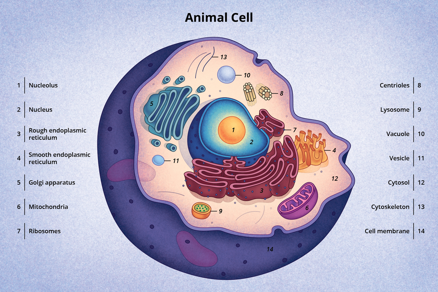
PDF Human Cell Diagram, Parts, Pictures, Structure and Functions The cell membraneis the outer coating of the cell and contains the cytoplasm, substances within it and the organelle. It is a double-layered membrane composed of proteins and lipids. The lipid molecules on the outer and inner part (lipid bilayer) allow it to selectively transport substances in and out of the cell. Endoplasmic Reticulum
Cell Membrane - The Definitive Guide | Biology Dictionary Definition. The cell membrane, also known as the plasma membrane, is a double layer of lipids and proteins that surrounds a cell. It separates the cytoplasm (the contents of the cell) from the external environment. It is a feature of all cells, both prokaryotic and eukaryotic. a 3D diagram of the cell membrane.
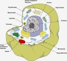
A Well-labelled Diagram Of Animal Cell With Explanation - BYJUS Well-Labelled Diagram of Animal Cell The Cell Organelles are membrane-bound, present within the cells. There are various organelles present within the cell and are classified into three categories based on the presence or absence of membrane. Listed below are the Cell Organelles of an animal cell along with their functions.
Cell Membrane Diagram Labeled : Functions and Diagram - ACTUINDE Cell Membrane Diagram Labeled Monday, March 22nd 2021. | Diagram Cell Membrane Diagram. There are no organelles in the prokaryotic cells, i.e., they have no internal membrane systems. While lipids help to give membranes their flexibility, proteins monitor and maintain.
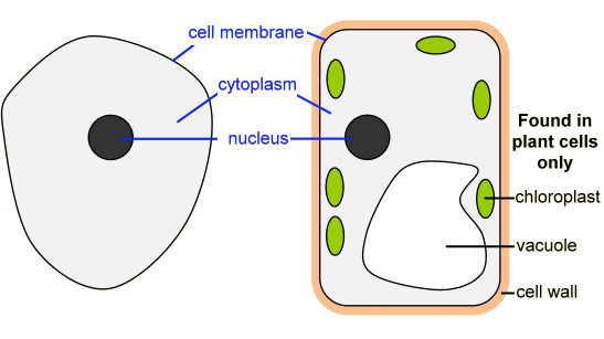
Plant Cell: Diagram, Types and Functions - Embibe Exams It is located outside the cell membrane and is completely permeable. The primary function of a plant cell wall is to protect the cell against mechanical stress and to provide a definite form and structure to the cell. ... The plant cell diagram can be checked above and on a similar pattern the diagram can be created. Q.3. Why do plant cells ...

Explain the nucleus of a cell with a neat labeled diagram - Science - Cell - Structure and ...
PDF Membrane Structure and Function - Phoenix College Major Components of the Cell Membrane: Lipids • Phospholipids are amphipathicmolecules (with hydrophobictails and a hydrophilichead) • One of the phospholipid tails exist mostly in a transconfiguration, providing more fluidityto the membrane • Cholesterol is a rigid molecule that makes membranes less fluid Cholesterol
Animal Cells: Labelled Diagram, Definitions, and Structure - Research Tweet The endoplasmic reticulum (s) are organelles that create a network of membranes that transport substances around the cell. They have phospholipid bilayers. There are two types of ER: the rough ER, and the smooth ER. The rough endoplasmic reticulum is rough because it has ribosomes (which is explained below) attached to it.
Cell Membrane Functions, Structure and Diagram - Jotscroll The Cell membrane or Plasma membrane is a thin selectively permeable layer that separates the interior of the cell from its outside environment. The cell membrane serves as the outer boundary of a living cell and also forms a boundary for an internal cell compartment that encloses organelles.
Structure of Membrane in Cells (With Diagram) - Biology Discussion Cell walls are formed through apposition, i.e., the wall material is deposited by the protoplast on the plasma membrane. The first cell wall (Primary wall) is formed during the cell growth phase. When the cell elongation is stopped the secondary cell wall, formation starts (Fig. 2.13).
Plant Cells: Labelled Diagram, Definitions, and Structure - Research Tweet Plastids and Chloroplasts. Plants make their own food through photosynthesis. Plant cells have plastids, which animal cells don't. Plastids are organelles used to make and store needed compounds. Chloroplasts are the most important of plastids. They convert light energy from the sun into sugar and oxygen. The most exposed parts of the plants ...
CELL MEMBRANE LABEL Diagram | Quizlet Practice labeling the parts of the cell membrane Terms in this set (6) Channel Protein hole or tunnel that particles may pass through to go in / out of cell Marker protein identifies or labels the cell Receptor protein receives information Heads part of the phospholipid that loves water (hydrophili) - points to the most outside and inside of cell


Post a Comment for "45 cell membrane diagram with labels"