38 onion cells under microscope with labels
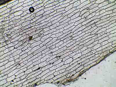
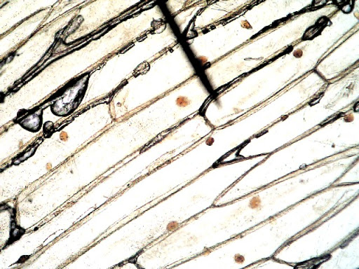
Onion Skin Cells - Investigation - Exploring Nature Observe the onion tissue under the microscope at 4x, 10x and 40x with lots of light (open diaphragm). Then slowly close the diaphragm while observing the image to find the best light for seeing cellular details. 6. Draw a section of onion skin cells at 10x magnification. Then switch to 40x and draw one cell and label it. Cells and Reproduction - BBC Bitesize All living organisms are made up of cells. Cells are the building units of life - the basic building blocks of all animals and plants. They are so small, you need to use a light microscope to see ...
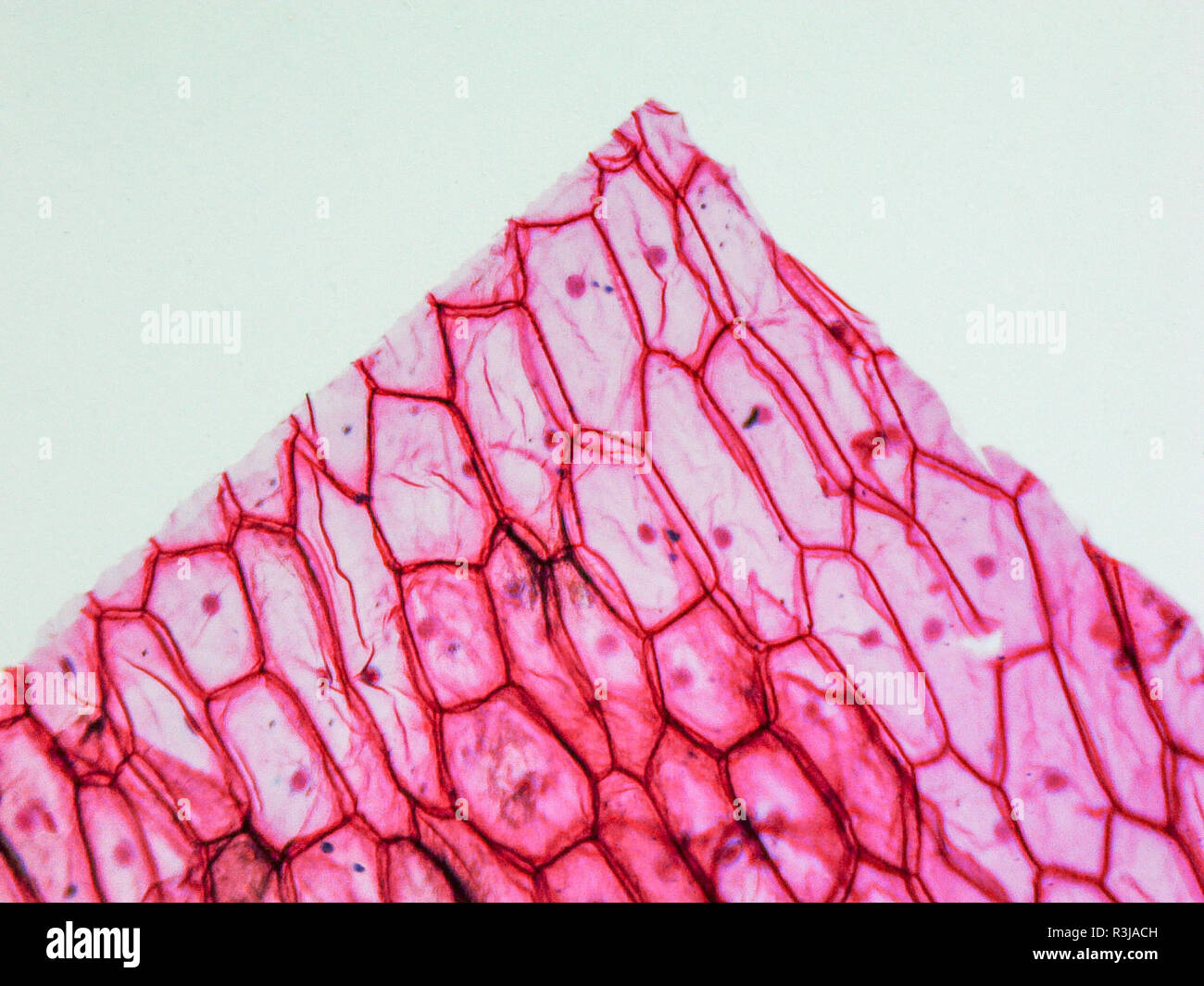
What organelles are in an onion cell? - Biology Stack Exchange You cannot see most of these as they appear translucent as well as being too small to see under the light microscope. You need an electron microscope to view these. Note: chloroplasts are not present in an onion cell as it is not a photosynthesising cell. This is a typical onion cell slide with labels:

Onion cells under microscope with labels
Onion Root Cells Dividing By Mitosis Under A Light Microscope ... - iStock Download this Onion Root Cells Dividing By Mitosis Under A Light Microscope At 100x Magnification Cells Visible In Prophase Metaphase Anaphase And Telophase photo now. And search more of iStock's library of royalty-free stock images that features Anaphase photos available for quick and easy download. Onion Peels Observed Under the Microscope | Confirmation Point Onion Peels Observed Under the Microscope Cells present in onion peel can be observed under microscope. For this onion peels are first isolated. For this experiment outer most scale of the onion is removed and is cut into four equal halves. It is a monocot plant. Then with the help of a pairs of forcep the scale of onion is peeled out. Cambridge International AS & A Level Biology Coursebook … 1.1 Cells are the basic units of life 1.2 Cell biology and microscopy 1.3 Plant and animal cells as seen with a light microscope 1.4 Measuring size and calculating magnification 1.5 Electron microscopy 1.6 Plant and animal cells as seen with an electron microscope 1.7 Bacteria 1.8 Comparing prokaryotic cells with eukaryotic cells 1.9 Viruses
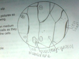
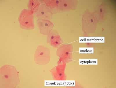
Onion cells under microscope with labels. Onion Cell Under Microscope Labeled a scientist is observing onion cells and human cheek cells under a microscope label the cell wall, nucleus, and cytoplasm draw and label your observations: label the nucleus, cell wall, and cytoplasm of one onion cell students know the characteristics that distinguish plant cells from animal cells, including chloroplasts and cell walls once you … Cambridge International AS and A Level Biology ... - Academia.edu BIO1: Maintaining a Balance 1. Most organisms are active in a limited temperature range IDENTIFY THE ROLE OF ENZYMES IN METABOLISM, DESCRIBE THEIR CHEMICAL COMPOSITION AND USE A SIMPLE MODEL TO DESCRIBE … Onion Cell Lab Report.docx - Onion Cell Lab Report By station, remove the single layer of epidermal cells from inner side of the scale leaf. 3(Place the single layer of onion on a glass slide. 4(Place a drop of iodine stain on your onion tissue. 5(Put the cover slip on the stained tissue and gently tap out any air bubbles. 6(Observe the cells under the microscope and see you results. Onion Root Cell Cycle Lab Answers | SchoolWorkHelper The stage that the cell is currently in is prophase. Also, the cell walls in the onion root were barely visible, but the nuclei were very clear. This was all seen in 400X total magnification. When observing the whitefish embryo cells for the stage of metaphase, the cells took on a circular shape and, like the onion root cell, many nuclei can be ...
The Biology Project The Biology Project, an interactive online resource for learning biology developed at The University of Arizona. The Biology Project is fun, richly illustrated, and tested on 1000s of students. It has been designed for biology students at the college and high school level, but is useful for medical students, physicians, science writers, and all types of interested people. DiFiore's Atlas of Histology with Functional Correlations (11th … Atlas of Human Histology A Guide to Microscopic Structure of Cells, Tissues and Organs. by Растко Корњача. Download Free PDF Download PDF Download Free PDF View PDF. The Eye and Ear: Special Sense Organs Appendix: Light Microscopy Stains. by bub bub. Download Free PDF Download PDF Download Free PDF View PDF. Download Download PDF. Download Full … Onion Root Tip Mitosis - Stages, Experiment and Results - MicroscopeMaster · Place a cap/lid onto the vial (ensure that the cap/lid has a pinprick hole) and place the vial in the water bath (at 55 degrees C) for about 5 minutes - This enhances the staining process · Using the forceps, remove the root tips from the vial of stain and place them onto a clean microscope glass slide Nano based drug delivery systems: recent developments and … 19.09.2018 · Recently, there has been enormous developments in the field of delivery systems to provide therapeutic agents or natural based active compounds to its target location for treatment of various aliments [33, 34].There are a number of drug delivery systems successfully employed in the recent times, however there are still certain challenges that need to be addresses and an …
ASSIST | Australian school science information support for … Science ASSIST has an expert national advisory team with extensive, collective experience across all school laboratory management and safety. It is this team that will help with your enquiry. Animal Cell Mitosis Under Microscope - Casey Sillman The division of the cell in two (cytokinesis) occurs chromosomes decondense (no longer visible under light microscope). In cell biology, mitosis (/maɪˈtoʊsɪs/) is a part of the cell cycle in which replicated chromosomes are separated into two new nuclei. Plant cells do not have centrioles like animal cells, just centrosomes. ONION CELLS VIDEO - YouTube Video shows how to make a wet mount slide to view onion cells under the microscope. Onion Cells Microscope Stock Photos and Images - Alamy Onion Cells under the Microscope ID: FC0CWP (RF) Onion skin cells under the microscope, horizontal field of view is about 0.61 mm ID: 2AM97C0 (RM) Detailed view of the cells of a red onion as seen through a microscope. Biology experiment. ID: 2H3C0XC (RF) Onion cells under a light microscope at 10 times magnification ID: 2EW4298 (RF)

Life Science Unit - WELCOME TO MR.FLEMING SCIENCE
Onion cells hi-res stock photography and images - Alamy RF 2BN75YG - Under the microscope onion cells. RF MRGPTT - Onion epidermis under light microscope. Purple colored, large epidermal cells of an onion, Allium cepa, in a single layer. Photo. RM EBXPHH - Cell walls and organelles of onion bulb scale epidermis cells. RM CP074T - mitosis in a onion root, anaphasis.

Biology Pictures: Onion Cells under Microscope | Biology, Cell biology, Picture
PDF Onion Cells - Investigation - Exploring Nature 5. Observe the onion tissue under the microscope at 4x, 10x and 40x with lots of light (open diaphragm). Then slowly close the diaphragm while observing the image to find the best light for seeing cellular details. 6. Draw a section of onion skin cells at 10x magnification. Then switch to 40x and draw one cell and label it. Questions: 1.

Cell Biology Project - Parts of a cell - Quatr.us Study Guides
Observing Cork Cells Under The Microscope » Microscope Club Place the cork dust on the microscope slide with a drop of water, then add another water droplet on top of the cork sample. Cover the prepared slide with a cover slip. Method 2 Alternatively, slice thin cork slices, making sure that ample light can pass through the slice, allowing you to see the cell layout and the individual cells.

Cryogenic scanning electron micrograph of the interface | Open-i
Observing Onion Cells Under The Microscope » Microscope Club Afterwards, carefully mount the prepared and stained onion cell slide onto the microscope stage. Make sure that the cover slip is perfectly aligned with the microscope slide, and that any excess stain has been wiped off. Secure the slide on the stage using the stage clips.

My Three Seeds of Joy Homeschool: Discovering Cells
Plant Cell Under Microscope 40X / Plant Cells Under Microscope 400x ... Assignment 6 Page 2 from projects.ncsu.edu Label the cell wall, cytoplasm (cyto = cell). Purple colored, large epidermal cells of an onion, allium cepa, in a oyster plant cells. ... This section on microscopy is meant as an introduction as learners will need to be able to use these same onion cells were viewed under a microscope which had not ...
General Biology Microscopic Specimen Images & Photographs
Onion Epidermis - kuensting.org Onion epidermal cells, iodine stain, 400X. The nucleus of an onion epidermal cell, 1000X magnification. ...
)
Onion Cells Under Microscope Stock Footage Video 4946132 | Shutterstock
Plant Cell Under Microscope 40X Labeled - Barrett Orey Set up your microscope, place the onion root slide on the stage and focus on low (40x) power. 3) to draw and label a plant cell under 40x, a spider under 4x and human blood under 100x objective lens. Compare animal and plant cells and distinguish each type under the microscope.
My Personal Experience - SusannaLaRochellemicroscopy
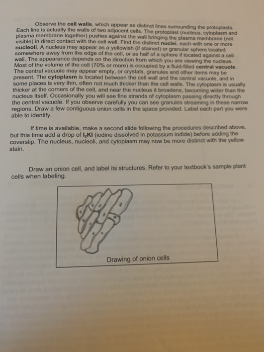
DOC Plant and Animal Cells Microscope Lab - hillsboro.k12.oh.us Make a drawing of one onion cell, labeling all of its parts as you observe them. (At minimum you should observe the nucleus, cell wall, and cytoplasm.) Cheek cells 1. To view cheek cells, gently scrape the inside lining of your cheek with a toothpick. DO NOT GOUGE THE INSIDE OF YOUR CHEEK! (We will observe blood cells in a future lab!!) 2.

The Microscope
Your liver is essential to your life. The Canadian Liver Foundation Because of its wide-ranging responsibilities, your healthy liver can come under attack by viruses, toxic substances, contaminants and diseases. However, even when under siege, the liver is very slow to complain. People who have problems with their liver are frequently unaware because they may have few, if any, symptoms. Your liver is such a determined organ that it will continue …

Onion Leaf Cell Under Microscope - Micropedia
Structure and function of mitochondrial membrane protein … 29.10.2015 · Mitochondria can be seen in the light microscope, but their detailed internal structure is only revealed by electron microscopy. In the 1990s, the structure of mitochondria was investigated by electron tomography of thin plastic sections . While this yielded striking three-dimensional (3D) images of their internal membrane system, molecular detail was lost due to …
Onion Cells Microscope High Resolution Stock Photography and Images - Alamy
American Express Prepare a wet mount with those cells. Remove the skin of the provided onion and carefully slice a small specimen to observe. Prepare it in a wet mount. B) 1. Observe and draw your skin cells (wet mount) at the frequency: 10X. 50X. 100X. 2. Observe and draw the prepared wet mount containing the onion cells at the frequency: 10X. 50X. 100X. 3.

Sketch the onion peel cell as seen under the microscope. Label the parts such as the cell wall ...
Microscope Cell Lab: Cheek, Onion, Zebrina | SchoolWorkHelper The first lab exercise was observing animal cells, in this case, my cheek cells. The second lab exercise was observing plant cells, in this case, onion epidermis. The third lab exercise was observing chloroplasts and biological crystals, in this case, a thin section from the Zebrina plant. The first thing that was done in this lab exercise was ...
Microscope Onion Cell Labeled - Micropedia
Onion Plant Cell Under Microscope Labeled - Ismael Dauila Explore diffusion/osmosis by looking at onion cells under the microscope. It is used for treating a parasite disease called ich (ichthyophthirius multifiliis; Label the cell wall and chloroplasts. Students will observe plant cells using a light microscope.

Onion Cell Under Microscope 40x Labeled - Micropedia
Vii sketch the onion peel cell as seen under the - Course Hero 15 Biology Practical BIOLOGY Notes Biology Practical 2.1.5 FOR THE TEACHER Please ensure that 1. slides and coverslips are cleaned before use. 2. microscope is handled properly. 3. staining is properly done as staining helps to highlight certain cell components. 4. the students are told that cell has other components also but they can not be seen under compound microscope.

Scientific Videos: Hydrilla: Hydrilla Leaf Cells
Plant Cell Under Microscope Observation : Grass cells under a ... Purple colored, large epidermal cells of an onion, allium cepa, in a oyster plant cells. Your plant cells under microscope stock images are ready. Observe the slide under microscope. Observe the labeled diagram of plant cell. Draw the structure that you see under microscope!! Place the glass slide onto the stage.
Biology Pictures: Onion Cells under Microscope
Onion Cells Under a Microscope (100x-2500x) - YouTube 567 views Feb 24, 2021 In this video you will see onion cells under a microscope (100x-2500x) as is, without any coloring. To observe the onion cells the thin membrane is used. It can easil ...more...
Plant & Animal Cells Staining Lab Answers | Online Homework Help | SchoolWorkHelper
Onion Cell Diagram Labeled Pdf [PDF] - thesource2.metro Set your multimeter to measure current in the 20 mA range (the dial setting labeled "20m" on the right). Plug the multimeter's black probe into the port labeled COM. Plug the multimeter's red probe into the port labeled VΩmA. Use a red alligator clip lead to connect the multimeter's red probe to the positive (+) terminal of the 9 V battery.

Fanos' MCB Blog
Onion Cells Under a Microscope - Requirements/Preparation/Observation Add a drop of iodine solution on the onion membrane (or methylene blue) Gently lay a microscopic cover slip on the membrane and press it down gently using a needle to remove air bubbles. Touch a blotting paper on one side of the slide to drain excess iodine/water solution, Place the slide on the microscope stage under low power to observe.
Post a Comment for "38 onion cells under microscope with labels"