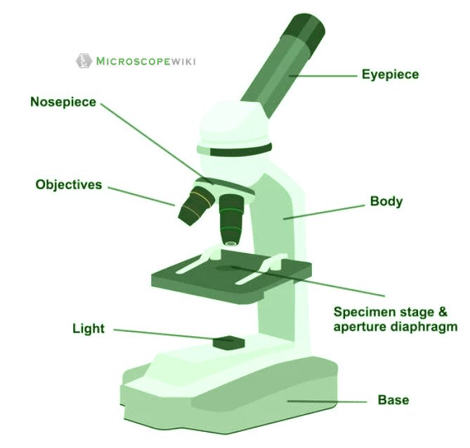
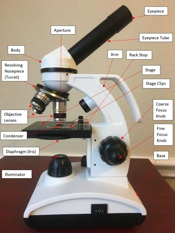
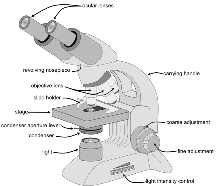
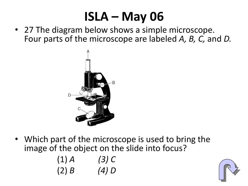
43 simple microscope diagram with labels
The Parts of a Microscope (Labeled) Printable Printable (6th - 12th ... The Parts of a Microscope (Labeled) Printable. Download. Add to Favorites. Share. This diagram labels and explains the function of each part of a microscope. Use this printable as a handout or transparency to help prepare students for working with laboratory equipment. Grade: Marine ecosystem — Science Learning Hub Clicking on the labels will bring up short video and images that can be used to explore marine ecosystems in greater detail. Use this interactive with the article Marine food webs . Excellent interactive for all, it would work brilliantly for all learners regardless of all reading abilities!
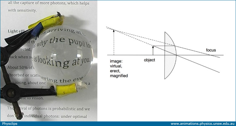
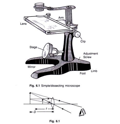
Simple Microscope - Diagram (Parts labelled), Principle, Formula and Uses Simple microscope is a magnification apparatus that uses a combination of double convex lens to form an enlarged, erect image of a specimen. The working principle of a simple microscope is that when a lens is held close to the eye, a virtual, magnified and erect image of a specimen is formed at the least possible distance from which a human eye ...
Simple microscope diagram with labels
Electron microscope - Wikipedia An electron microscope is a microscope that uses a beam of accelerated electrons as a source of illumination. As the wavelength of an electron can be up to 100,000 times shorter than that of visible light photons , electron microscopes have a higher resolving power than light microscopes and can reveal the structure of smaller objects. Simple Microscope: Definition, Principle, Parts, And Uses A simple microscope is a rudimentary magnification device that is capable of visibly enlarging small objects, so they can be viewed and studied in better detail. It was invented in the late 16th century, and is still being widely used today. Simple microscopes have a wide range of applications in various fields. Microscope Labeling - The Biology Corner Students label the parts of the microscope in this photo of a basic laboratory light microscope. Can be used for practice or as a quiz. ... The type of microscope used in most science classes is the _____ microscope. 18. You should carry the microscope by the _____ and the _____. 19. The objectives are attached to what part of the microscope ...
Simple microscope diagram with labels. Achiever Papers - We help students improve their academic ... Professional academic writers. Our global writing staff includes experienced ENL & ESL academic writers in a variety of disciplines. This lets us find the most appropriate writer for any type of assignment. Microscope labeled diagram - SlideShare Microscope labeled diagram 1. The Microscope Image courtesy of: Microscopehelp.com Basic rules to using the microscope 1. You should always carry a microscope with two hands, one on the arm and the other under the base. 2. You should always start on the lowest power objective lens and should always leave the microscope on the low power lens ... Simple Microscope - Parts, Functions, Diagram and Labelling Parts of the optical parts are as follows: Mirror - A simple microscope has a plano-convex mirror and its primary function is to focus the surrounding light on the object being examined. Lens - The biconvex lens is placed above the stage and its function is to magnify the size of the object being examined. Simple Microscope - Definition, Types, Working Principle & Formula 1. Simple microscope comprises a biconvex lens used as a magnifying glass. Compound microscope comprises 2 or more convex lenses where one lens is the eyepiece and the other one is the objective lens. 2. Natural light is the source to see the object. An illuminator is a source to see the object. 3.
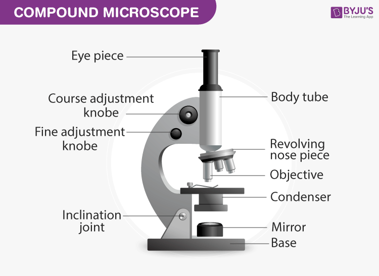
Microscope Drawing Easy with Label - YouTube In this video I go over a microscope drawing that is easy with label. There is a blank copy at the end of the video to review on your own. A great way to s... Microscope, Microscope Parts, Labeled Diagram, and Functions Revolving Nosepiece or Turret: Turret is the part of the microscope that holds two or multiple objective lenses and helps to rotate objective lenses and also helps to easily change power. Objective Lenses: Three are 3 or 4 objective lenses on a microscope. The objective lenses almost always consist of 4x, 10x, 40x and 100x powers. The most common eyepiece lens is 10x and when it coupled with ... Compound Microscope Parts - Labeled Diagram and their Functions The term "compound" refers to the microscope having more than one lens. Basically, compound microscopes generate magnified images through an aligned pair of the objective lens and the ocular lens. In contrast, "simple microscopes" have only one convex lens and function more like glass magnifiers. [In this figure] Two "antique ... Microscope Poster - Diagram with Labels | Teach Starter A poster containing a diagram with labels showing the key parts of a microscope. In Science it is important that students know how to use a variety of tools when conducting scientific experiments and inquiry. This poster focuses on the microscope and highlights its key parts. Print on tabloid paper to display around your school's science lab ...
Label the Microscope Diagram | Download Scientific Diagram - ResearchGate Gram staining was performed using compound microscope according to the procedure described by Petersen et al., 2016 [8]. The gram positive and gram negative bacteria were identified based on ... Parts of a Microscope Labeling Activity - Storyboard That Knowing the names of the different parts of the microscope is essential to be able to use one properly. Create a poster that labels the parts of a microscope and includes descriptions of what each part does. Click "Start Assignment". Use a landscape poster layout (large or small). Search for a diagram of a microscope. Label the microscope — Science Learning Hub In this interactive, you can label the different parts of a microscope. Use this with the ... A Study of the Microscope and its Functions With a Labeled Diagram ... To better understand the structure and function of a microscope, we need to take a look at the labeled microscope diagrams of the compound and electron microscope. These diagrams clearly explain the functioning of the microscopes along with their respective parts. Man's curiosity has led to great inventions. The microscope is one of them.
Microscope With Labels clip art | Microscope parts, Scientific method ... Description Use this blank handout as a way for students to record microscope drawings. Aside from the drawig itself, studnts are prompted to title the drawing, include the magnification of the microscope, and give a quick description of what they are viewing. Keywords: Microscope Drawings Lab Biology. S. Learning with Shedd Aquarium.
Morphological diversity of single neurons in molecularly ... Oct 06, 2021 · a, Example single-neuron morphologies for each of the 11 projection neuron types.Numbers in parentheses denote the number of neurons shown in each indicated region. In this and all subsequent ...
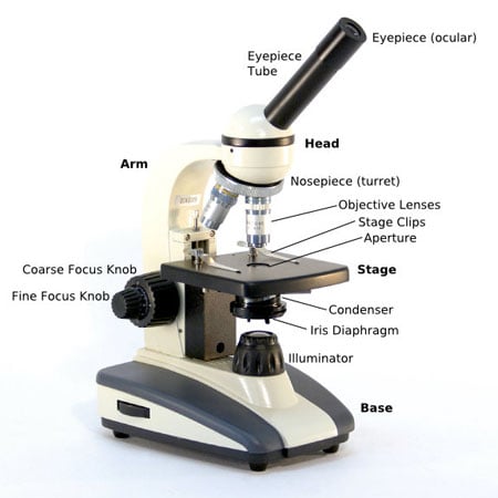
16 Parts of a Compound Microscope: Diagrams and Video Once you have an understanding of the parts of the microscope it will be much easier to navigate around and begin observing your specimen, which is the fun part! The 16 core parts of a compound microscope are: Head (Body) Arm. Base. Eyepiece. Eyepiece tube.
A Study of the Microscope and its Functions With a Labeled Diagram ... May 21, 2019 - To better understand the structure and function of a microscope, we need to take a look at the labeled microscope diagrams of the compound and electron microscope. These diagrams clearly explain the functioning of the microscopes along with their respective parts.
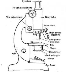
Labelled Diagram of Compound Microscope The below mentioned article provides a labelled diagram of compound microscope. Part # 1. The Stand: The stand is made up of a heavy foot which carries a curved inclinable limb or arm bearing the body tube. The foot is generally horse shoe-shaped structure (Fig. 2) which rests on table top or any other surface on which the microscope in kept.
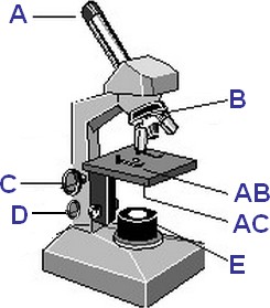
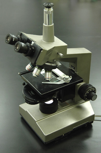
Microscope Labeling - The Biology Corner Microscope Labeling. Shannan Muskopf May 31, 2018. This simple worksheet pairs with a lesson on the light microscope, where beginning biology students learn the parts of the light microscope and the steps needed to focus a slide under high power. The labeling worksheet could be used as a quiz or as part of direct instruction where students label the microscope as you go over what each part is used for.
Simple Microscope- Definition, Principle, Magnification, Parts ... A simple microscope is one that uses a single lens for magnification, such as a magnifying glass while a compound microscope uses several lenses to enhance the magnification of an object. It uses a lens to enlarge an object through angular magnification alone, giving the viewer an erect enlarged virtual image. The use of a single convex lens or ...
Microscope Types (with labeled diagrams) and Functions Simple microscope labeled diagram. Simple microscope functions. It is used in industrial applications like: Watchmakers to assemble watches; Cloth industry to count the number of threads or fibers in a cloth; Jewelers to examine the finer parts of jewelry; Miniature artists to examine and build their work; Also used to inspect finer details on products
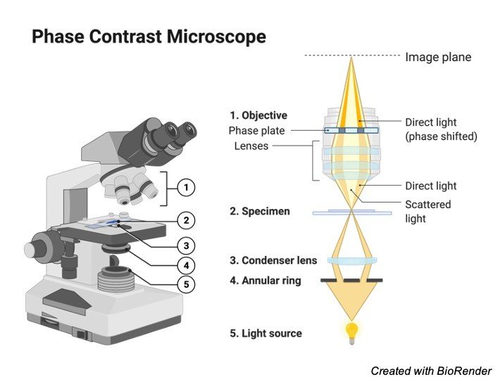
Parts of a microscope with functions and labeled diagram - Microbe Notes Head - This is also known as the body. It carries the optical parts in the upper part of the microscope. Base - It acts as microscopes support. It also carries microscopic illuminators. Arms - This is the part connecting the base and to the head and the eyepiece tube to the base of the microscope.
Simple Microscope Definition, Magnification, Parts And Uses - BYJUS Following are the parts of the simple microscope with their functions: Eyepiece: It is the lens that is used to study the samples and is placed at the top. It has a magnification of 10X to 15X. Base: This provides support to the microscope. Tube: This is used to connect the eyepiece to the objective lenses.
Parts of the Microscope with Labeling (also Free Printouts) 5. Knobs (fine and coarse) By adjusting the knob, you can adjust the focus of the microscope. The majority of the microscope models today have the knobs mounted on the same part of the device. Image 5: The circled parts of the microscope are the fine and coarse adjustment knobs. Picture Source: bp.blogspot.com.
Success Essays - Assisting students with assignments online Get 24⁄7 customer support help when you place a homework help service order with us. We will guide you on how to place your essay help, proofreading and editing your draft – fixing the grammar, spelling, or formatting of your paper easily and cheaply.
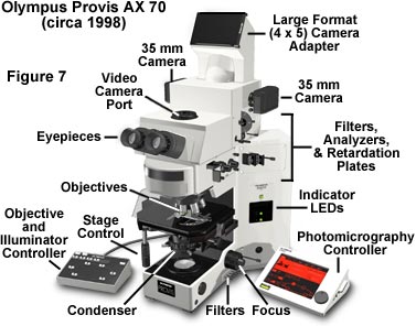
Compound Microscope Parts, Functions, and Labeled Diagram Compound Microscope Definitions for Labels. Eyepiece (ocular lens) with or without Pointer: The part that is looked through at the top of the compound microscope. Eyepieces typically have a magnification between 5x & 30x. Monocular or Binocular Head: Structural support that holds & connects the eyepieces to the objective lenses.
Free Microscope Worksheets for Simple Science Fun for Your Students 1. Parts of a Microscope . The first worksheet labels the different parts of a microscope, including the base, slide holder, and condenser. If you have a microscope, compare and contrast this worksheet to it.Also, your kids can color this microscope diagram in and read the words to each part of the microscope.
Parts of a Simple Microscope - Labeled (with diagrams) image 5: A modern simple microscope with the different parts labeled. image source: laboratoryinfo.com The optical parts of a simple microscope are centered on the specimen - lighting, and magnification.
Frontiers | Virus Detection: A Review of the Current and ... Apr 20, 2021 · Viruses are ubiquitous in the environment. While many impart no deleterious effects on their hosts, several are major pathogens. This risk of pathogenicity, alongside the fact that many viruses can rapidly mutate highlights the need for suitable, rapid diagnostic measures. This review provides a critical analysis of widely used methods and examines their advantages and limitations. Currently ...
Spin-enhanced nanodiamond biosensing for ultrasensitive ... Nov 25, 2020 · c, A microscope image of the omega-shaped stripline resonator used to produce a uniform field at 2.87 GHz. d, Excitation and emission spectra of FNDs. The green shaded area shows the filtered ...
Simple Squamous Epithelium under a Microscope with a Labeled Diagram ... Simple Squamous Epithelium under a Microscope with a Labeled Diagram. 04/04/2022 03/04/2022 by anatomylearner. Simple squamous epithelium under a microscope consists of a single layer of thin, flat, and scale-like cells. These cells are joined together by an intercellular junction and rest on the basement membrane, whose thickness depends on the location.
Microscope Labeling - The Biology Corner Students label the parts of the microscope in this photo of a basic laboratory light microscope. Can be used for practice or as a quiz. ... The type of microscope used in most science classes is the _____ microscope. 18. You should carry the microscope by the _____ and the _____. 19. The objectives are attached to what part of the microscope ...
Simple Microscope: Definition, Principle, Parts, And Uses A simple microscope is a rudimentary magnification device that is capable of visibly enlarging small objects, so they can be viewed and studied in better detail. It was invented in the late 16th century, and is still being widely used today. Simple microscopes have a wide range of applications in various fields.
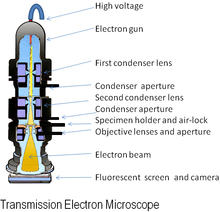
Electron microscope - Wikipedia An electron microscope is a microscope that uses a beam of accelerated electrons as a source of illumination. As the wavelength of an electron can be up to 100,000 times shorter than that of visible light photons , electron microscopes have a higher resolving power than light microscopes and can reveal the structure of smaller objects.















Post a Comment for "43 simple microscope diagram with labels"